3.5.5
3 - 47
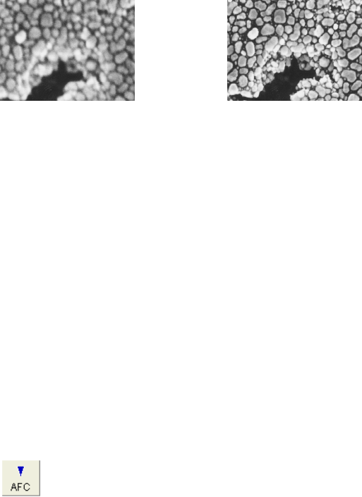
(b) Adjust the stigmators X and Y alternately for the sharpest image.
Fig. 3.5-19 Stigma Adjustment
(c) Focus again and check image drift and sharpness.
(d) Repeat steps (a) to (c) until adjustments are completed.
NOTICE: If it takes a long time to focus and correct astigmatism, you may end up with specimen
damage due to electron beam irradiation and/or contamination.
If the specimen is beam- or contamination-sensitive, we suggest the following
techniques:
(a) Reduce probe current.
(b) Use another area on the specimen for focusing purposes. After focusing, return
to the area of interest, adjust the final focus quickly, and then capture or record
the image.
(2) Auto Focus function
Click Auto button
on the Control panel or select the Auto Focus command from the
Operate menu to start Auto Focus.
When magnification is lower than 5,000×, coarse focus (search using a wide focus range) is
carried out. Fine focus (search using a narrow focus range) is carried out at magnifications
higher than 5,000×.
Fine focus works correctly under conditions where the image is not clear but visible.
The result of Auto Focus depends on the surface structures of the specimen. When there
is little or no surface detail on the specimen, or when the specimen is charged, Auto Focus
does not operate properly.


