


ECG Lead Placements 6 ECG, Arrhythmia, and ST Monitoring
99
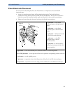
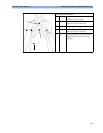
Chest Electrode Placement
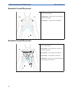
For accurate chest electrode placement and measurement, it is important to locate the fourth
intercostal space.
1 Locate the second intercostal space by first palpating the Angle of Lewis (the little bony
protuberance where the body of the sternum joins the manubrium). This rise in the sternum is
where the second rib is attached, and the space just below this is the second intercostal space.
2 Palpate and count down the chest until you locate the fourth intercostal space.
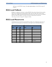
V1 placement: on the fourth
intercostal space at the right sternal
border
V2 placement: on the fourth
intercostal space at the left sternal
border
V3 placement: midway between the
V2 and V4 electrode positions
V4 placement: on the fifth
intercostal space at the left
midclavicular line
V5 placement: on the left anterior
axillary line, horizontal with the V4
electrode position
V6 placement: on the left midaxillary line, horizontal with the V4 electrode position
V3R to V6R placement: on the right side of the chest in positions corresponding to those on the left
VE placement: over the xiphoid process
V7 placement: on posterior chest at the left posterior axillary line in the fifth intercostal space
V7R placement: on posterior chest at the right posterior axillary line in the fifth intercostal space
VE
V1
V2
V3
V4
V5
V6
V7
V3R
V4R
2
3
4
Angle of
Lewis