


69
7. Start-up
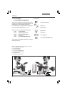
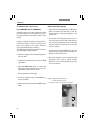
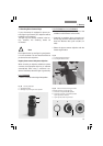
• Using the condenser height adjuster (90.2)
, ad-
just the condenser until the edge of the field
diaphragm appears in sharp relief (not S70
condenser).
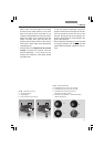
• Open the field diaphragm until
it only just disappears from the field of view
(91d).
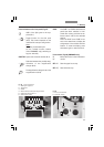
Leica DMI4000B and Leica DMI6000B:
• Select an objective with moderate magnifica-
tion (10x–20x).
• Activate the transmitted-light axis with the
TL/IL button (84.1).
• Press the BF button to activate the bright field
contrast method (one of the variable function
buttons on the stand).
• Place a specimen on the stage.
• Focus the specimen using the SmartMove or
the focus wheels.
• Adjust the light intensity with the INT buttons
(84.3).
• Close the field diaphragm with the FD button
(84.4) or manually until the edge of the dia-
phragm appears in the field of view.
• Using the condenser height adjuster (90.2),
adjust the condenser until the edge of the
field diaphragm appears in sharp relief (not
S70 condenser).
• Open the field diaphragm just enough for it to
disappear from the field of view (91d).
Note:
The condenser height setting is dependent on
the thickness of the specimen and may require
adjustment for each new specimen.
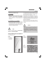
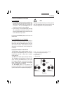
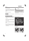
Koehler illumination
(not for S70 condenser)
Suitable values for the motorized aperture dia-
phragm and motorized field diaphragm have
been preset for each objective
(Leica DMI4000B
and Leica DMI6000B).
The condenser has also
been centered at the factory.
However, it may be necessary to readjust the
condenser in some cases. Therefore, check the
condenser centering.
The following procedure is provided for the
transmitted light-bright field illumination.
All required functions can be executed at the
touch of a button with the Leica DMI6000 elec-
tronic microscope. (See Chapter 8, Operation).
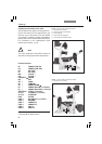
Preparation:
• Configure the microscope as follows:
Set up the illumination, condenser, objectives
and eyepieces correctly. (Please ensure that
the objectives are properly screwed in and
check the eyepiece settings.)
• Switch the microscope on and wait for the ini-
tialization phase to complete (automatic func-
tions only).
•You will need either an empty Petri dish (pref-
erably with a glass bottom) with a marking in
the middle or a stained specimen on a slide
with a coverslip.