


116
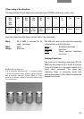
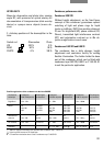
Performance data of eyepieces
Leica eyepiece Magnification/ Eyepiece port +)
type fov
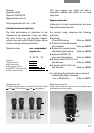
HC PLAN 10x/20
M
HC PLAN 10x/20
HC PLAN 12.5x/16 M
HC PLAN 10x/20 MF
HC PLAN 10x/22 M
HC PLAN 11x/20 MF
Eyepiece tube diameter: 30 mm
+)
= With removable or push-back anti-glare protection
for use with or without eyeglasses.
M = Ajustable eyelens (dioptre compensation) and slot
for graticules of 26 mm diameter.
MF = With illuminated graticule.
The LEITZ PERIPLAN
®
eyepiece type may not be used! Earlier
L PLAN type eypieces may only be used with earlier type tubes
(before about 1988) without the HC engraving!
Eyepiece field of view
Each microscope configuration has a certain
eyepiece field of view (see below), e. g. 20,
which must not be exceeded. If the maximum
fov is exceeded there may be disturbing loss of
definition and/or vignetting at the edge of the
image, → following pages!
The eyepiece field of view (fov) designates the
diameter of the intermediate image in the
eyepiece in mm, i.e. the diameter of the circular
diaphragm that frames the image and that lies
inside the eyepiece. This fov is specified on the
eyepiece after the magnification, e. g. 10x/20.
For the Leica DM IRB microscope we recom-
mend fov 22.
The maximum eyepiece field of view of a
specific configuration is derived from the
following microscope data:
Field performance of the objectives
Field performance of the intermediate
module(s)
Tube field number
Condenser properties
The decisive value is always the smallest.
For example, if the intermediate modules only
permit a field of view of 20 mm, but the
objectives and tube 25 mm, only eyepieces up to
fov 20 can be used. Eyepieces with fov 25 can
lead to vignetting in this case.
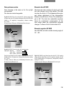
The diameter of the viewable specimen area is
calculated by dividing the diameter of the field
of view by the magnification of the objective and
the magnification factor of the microscope
optics.
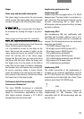
Example:
Eyepiece 10x/20
Objective PLAN 4/0.10
Magnification factor of the Leica DM IRB
Microscope optics 1x
Viewable specimen area
20 mm
–––––––––
= Ø 5 mm
4 x 1
The total magnification of the microscope is
worked out by multiplying the eyepiece mag-
nification with the reproduction ratio of the
objective and the magnification factor of the
microscope optics.