


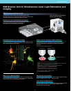
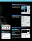


Spectral Based Detection
Flexibility and High Sensitivity
Spectral detection using gratings
for 2 nm wavelength resolution
and image acquisition matched to
fluorescence wavelength peaks.
User adjustable bandwidth of
emission spectrum for acquiring
bright images with minimal cross-
talk.
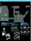
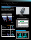
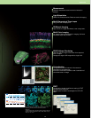
Precise Spectral Imaging
The spectral detection unit uses a grating method that offers
linear dispersion compared with prism dispersion. The unit
provides 2 nm wavelength resolution to high-sensitivity
photomultiplier tube detectors. Fluorescence separation can be
achieved through unmixing, even when cross-talk is generated
by multiple fluorescent dyes with similar peaks.
496 500 504 508 512 516 520 524
Wavelength
Intensity
528 532 536 540 544 548 552
400
600
800
1,000
1,200
1,400
1,600
1,800
2,000
2,200
2,400
2,600
EGFP (dendrite) — EYFP (synapse)
XYλ
Wavelength detection range: 495 nm–561 nm in
2 nm steps
Excitation wavelength: 488 nm
Courtesy of: Dr. Shigeo Okabe
Department of Anatomy and Cell Biology,
Tokyo Medical and Dental University
EGFP–EYFP Fluorescence Separation
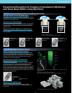
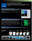
EYFP
EGFP
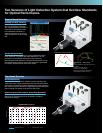
480
Conventional mirror unit High-performance mirror unit
500 520 540 560 580
Wavelength (nm)
600 620 640 660 680 700
0
20
40
Transmittance (%)
60
80
100
DM488/543/633 Comparison
Two Versions of Light Detection System that Set New Standards
for Optical Performance.
7
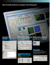
Filter Based Detection
Enhanced Sensitivity
Three-channel scan unit with detection system featuring hard
coated filter base. High-transmittance and high S/N ratio optical
performance is achieved through integration of a pupil projection
lens within the optics, the use of a high sensitivity photomultiplier
and an analog processing circuit with minimal noise.
High-Performance Filters Deliver Outstanding Separation
Special coatings deliver exceptionally sharp transitions to a
degree never achieved before, for acquisition of brighter
fluorescence images.