


Technology / Hardware
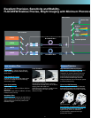
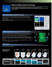
Switchable between Confocal and TIRFM Imaging
Switchable between confocal and TIRFM imaging for localization
of proteins on the cytoplasmic membrane surface and acquisition
of sectioning images within cells.
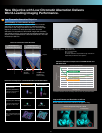
Software Control of TIRF Illumination
Built-in laser provides TIRF illumination. Software can be used to
tune the angle of incidence of excitation light and calculates the
penetration depth of the evanescent wave based on the TIRF
objective used.
High-Numerical Aperture Objectives for TIRF Illumination
A line of high-numerical aperture (NA) objectives is available for
TIRF illumination.
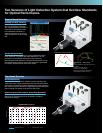
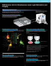
TIRFM (Total Internal Reflection Fluorescence Microscope) System
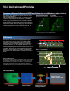
FV1000MPE Multiphoton Excitation System
Brighter and Deeper Imaging with Finer Resolution
The FV1000 is upgradeable to multiphoton excitation capability
by adding a dedicated laser and multiphoton optical system.
Optical design is optimized for multiphoton principles for brighter
imaging of features deep within living specimens, at higher
resolutions than previously possible.
Special Multiphoton Objective with Outstanding
Brightness and Resolution
Olympus offers a high NA water-immersion objective designed
for a wide field of view, with improved transmittance at near-
infrared wavelengths. A correction collar compensates for
spherical aberration caused by differences between the refractive
indices of water and specimens, forming the optimal focal spot
even in deep areas, without loss of energy density. The objective
is designed to collect scattered light over a wide field of view for
maximum image brightness.
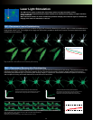
Multiphoton Laser Light Stimulation
Adding a multiphoton laser to the SIM scanner enables
multiphoton laser light stimulation or uncaging confined to the
focal volume.
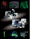
Exceptional Resolution for Imaging of Cytoplasmic Membrane
and Areas Deep Within Living Specimen.
GFP—Pak—K298A in HeLa cells.
Courtesy of Dr.J M Dong of sGSK-NRP laboratory, Singapore
LSMTIRFM
APON60xOTIRF
NA : 1.49 (oil immersion)
WD: 0.1 mm
Apo100xOHR
NA : 1.65 (oil immersion)
WD: 0.1 mm
(Customized cover glass and
immersion oil)
UAPON100xOTIRF
NA : 1.49 (oil immersion)
WD: 0.1 mm
UAPON150xOTIRF
NA : 1.45 (oil immersion)
WD: 0.08 mm
NEW
NEW
NEW
NEW
NEW
NEW
XLPLN25xWMP
Magnifications : 25x
NA : 1.05 (water immersion)
W.D. : 2.0 mm
3-dimensionally constructed images of neurons expressing EYFP in the cerebral neocortex of a
mouse under anesthesia.
Courtesy of:
Hiroaki Waki, Tomomi Nemoto, and Junichi Nabekura
National Institute for Physiological Sciences, National Institutes of Natural Sciences, Japan
* The FLUOVIEW FV1000MPE is a class 4 laser product.
10