


Objectives NA W.D. (mm) DIC prism Revolving
nosepiece
MPLN5X
0.10 20.00 –
WI-SSNP,
WI-SRE3
UMPLFLN10XW 0.30 3.50 WI-DIC10HR
WI-SSNP,
WI-SRE3
UMPLFLN20XW 0.50 3.50 WI-DIC20HR
WI-SSNP,
WI-SRE3
LUMPLFLN40XW 0.80 3.30 WI-DIC40HR
WI-SSNP,
WI-SRE3
LUMPLFLN60XW 1.00 2.00 WI-DIC60HR
WI-SSNP,
WI-SRE3
LUMFLN60XW 1.10 1.5 WI-DIC60HR
WI-SSNP,
WI-SRE3
XLUMPLFLN20XW 1.00 * 2.0 WI-DICXLU20HR WI-SNPXLU2
Objectives for fixed stage upright microscope
(using WI-UCD, WI-DICTHRA2)
* Note: These conditions are not met in confocal microscopy
W.D.
Cover glass
Correction
Condenser for BX2 Condenser for IX2
U-DICTS
Description NA thickness Immersion U-UCD8A-2 IX2-LWUCDA2
(mm)
(mm)
ring
optical element optical element
position
UPLSAPO4X 0.16 13 —
UPLSAPO10X2 0.40 3.1 0.17 U-DIC10 IX2-DIC10 normal
UPLSAPO20X 0.75 0.6 0.17 U-DIC20 IX2-DIC20 normal
UPLSAPO20XO 0.85 0.17 — Oil U-DIC20 IX2-DIC20 normal
UPLSAPO40X2 0.95 0.18 0.11-0.23 _ U-DIC40 IX2-DIC40 normal
UPLSAPO60XO 1.35 0.15 0.17 Oil U-DIC60 IX2-DIC60 BFP1
UPLSAPO60XW 1.20 0.28 0.13-0.21 Water _ U-DIC60 IX2-DIC60 normal
UPLSAPO100XO 1.40 0.12 0.17 Oil U-DIC100 IX2-DIC100 normal
PLAPON60XO 1.42 0.15 0.17 Oil U-DIC60 IX2-DIC60 BFP1
PLAPON60XOSC 1.40 0.12 0.17 Oil U-DIC60 IX2-DIC60 BFP1
UPLFLN40XO 1.30 0.2 0.17 Oil U-DIC40 IX2-DIC40 BFP1
APON60XOTIRF 1.49 0.1 0.13-0.19 Oil _ U-DIC60 IX2-DIC60 BFP1
UAPON100XOTIRF 1.49 0.1 0.13-0.19 Oil _ U-DIC100 IX2-DIC100 normal
UAPON150XOTIRF 1.45 0.08 0.13-0.19 Oil _ U-DIC100 IX2-DIC100 normal
Apo100XOHR 1.65 0.1 0.15 Oil U-DIC100 IX2-DIC100 normal
Objectives for BX2 and IX2
(using U-UCD8A-2, IX2-LWUCDA2 and U-DICTS)
Main Specifications
25
Spectral Version Filter Version
Laser Light Ultraviolet/Visible Light Laser LD lasers: 405 nm: 50 mW, 440 nm: 25 mW, 473 nm: 15 mW, 559 nm: 15 mW, 635 nm, 20 mW
Multi-line Ar laser (458 nm, 488 nm, 515 nm, Total 30 mW), HeNe(G) laser (543 nm, 1 mW)
AOTF Laser Combiner Visible light laser platform with implemented AOTF system, Ultra-fast intensity modulation with individual laser lines, additional shutter control
Continuously variable (0.1%–100%, 0.1% increment), REX: Capable of laser intensity adjustment and laser wavelength selection for each region
Fiber Broadband type (400 nm–650 nm)
Scanning and Scanner Module Standard 3 laser ports, VIS – UV – IR
Detection Excitation dichromatic mirror turret, 6 position (High performance DMs and 20/80 half mirror), Dual galvanometer mirror scanner (X, Y)
Motorized optical port for fluorescence illumination and optional module adaptation, Adaptation to microscope fluorescence condenser
Detector Module Standard 3 confocal Channels (3 photomultiplier detectors) Standard 3 confocal Channels (3 photomultiplier detectors)
Additional optional output port light path available for optional units Additional optional output port light path available for optional units
6 position beamsplitter turrets with CH1 and CH2 6 position beamsplitter turrets with CH1 and CH2
CH1 and CH2 equipped with independent grating and slit for fast and CH1 to CH3 each with 6 position barrier filter turret
flexible spectral detection (High performance filters)
Selectable wavelength bandwidth: 1–100 nm
Wavelength resolution: 2 nm
Wavelength switching speed: 100 nm/msec
CH3 with 6 position barrier filter turret
Filters High performance sputtered filters, dichromatic mirrors and barrier filters
Scanning Method 2 galvanometer scanning mirrors
Scanning Modes Scanning speed: 512 x 512 (1.1 sec., 1.6 sec., 2.7 sec., 3.3 sec., 3.9 sec., 5.9 sec., 11.3 sec., 27.4 sec., 54.0 sec.)
256 x 256 bidirectional scanning (0.064 sec., 0.129 sec.)
X,Y,T,Z,λ X,Y,T,Z
Line scanning: Straight line with free orientation, free line, Point scanning Line scanning: Straight line with free orientation, free line, Point scanning
Photo Detection Method 2 detection modes: Analog integration and hybrid photon counting
Pinhole Single motorized pinhole Single motorized pinhole
pinhole diameter ø50–300 µm (1 µm step) pinhole diameter ø50–800 µm (1 µm step)
Field Number (NA) 18
Optical Zoom 1x–50x in 0.1x increment
Z-drive Integrated motorized focus module of the microscope, minimum increment 0.01 µm or 10 nm
Transmitted Light Module with integrated external transmitted light photomultiplier detector and 100 W Halogen lamp, motorized switching, fiber adaptation to microscope
Detector unit frame
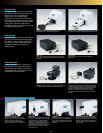
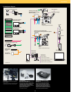
Microscope Motorized Microscope Inverted IX81, Upright BX61, Upright focusing nosepiece & fixed stage BX61WI
Fluorescence Illumination External fluorescence light source with motorized shutter, fiber adaptation to optical port of scan unit
Unit Motorized switching between LSM light path and fluorescence illumination
System Control PC PC-AT compatible, OS: Windows XP Professional (English version), Windows Vista (English version), Memory: 2.0 GB or larger, CPU:Core2Duo 3.0 GHz,
Hard disk: 500 GB or larger, Media: DVD Super Multi Drive, FV1000 Special I/F board (built-in PC), Graphic board: conformity with Open GL
Power Supply Unit Galvo control boards, scanning mirrors and gratings, Real time controller Galvo control boards, scanning mirrors
Display SXGA 1280X1024, dual 19 inch (or larger) monitors or WQUXGA 2560 x 1600, 29.8 inch monitor
Optional Unit SIM Scanner 2 galvanometer scanning mirrors, pupil projection lens, built-in laser shutter, 1 laser port, Fiber introduction of near UV diode laser or visible light laser,
Optional: 2nd AOTF laser combiner
TIRFM Unit Available laser: 405–633 nm. Motorized penetration ratio adjustment. Automatic optical setting for TIRFM objectives
4th CH Detector Module with photomultiplier detector, barrier filter turret, beamsplitter turret mounted with 3rd CH light path
Fiber Port for Fluorescence Output port equipped with FC fiber connector (compatible fiber core 100–125 µm)
Software
Image Acquisition Normal scan: 64 x 64, 128 x 128, 256 x 256, 320 x 320, 512 x 512, 640 x 640, 800 x 800, 1024 x 1024, 1600 x 1600, 2048 x 2048, 4096 x 4096
Clip rectangle scan ,Clip ellipse scan ,Polygon clip scan,line scan ,free line scan,Point scan, Real-time image
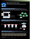
2-dimension: XY, XZ, XT and Xλ
3-dimension: XYZ, XYT, XYλ, XZT, XTλ and XZλ
4-dimension: XYZT, XZTλ and XYTλ
5−dimension: XYZTλ
Programmable Scan Controller Time Controller function
2D Image Display Each image display: Single-channel side-by-side, merge, cropping, live tiling, live tile, series (Z/T/λ),
LUT: individual color setting, pseudo-color, comment: graphic and text input
3D Visualization and Observation Interactive volume rendering: volume rendering display, projection display, animation displayed (save as OIF, AVI or MOV format)
Free orientation of cross section display
3D animation (maximum intensity projection method, SUM method)
3D and 2D sequential operation function
Image Format OIB/ OIF image format
8/ 16 bit gray scale/index color, 24/ 32/ 48 bit color,
JPEG/ BMP/ TIFF/ AVI/ MOV image functions
Olympus multi-tif format
Spectral Unmixing 2 Fluorescence spectral unmixing modes (normal and blind mode)
Image Processing Filter type: Sharpen, Average, DIC Sobel, Median, Shading, Laplacian
Calculations: inter-image, mathematical and logical, DIC background leveling
Image Analysis Fluorescence intensity, area and perimeter measurement, time-lapse measurement
Statistical Processing 2D data histogram display, colocalization
Optional Software Review station software, Off-line FLUOVIEW software for date analysis.
Motorized stage control software, Diffusion measurement package, Multi stimulation software, Multi area time-lapse software