


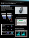
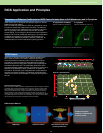
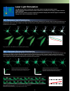
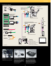

Laser Light Stimulation
The SIM scanner system combines the main scanner with a laser light stimulation scanner.
Control of the two independent beams enables simultaneous stimulation and imaging, to capture reactions
during stimulation.
Multi-stimulation software is used to continuously stimulate multiple points with laser light for simultaneous
imaging of the effects of stimulation on the cell.
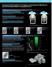
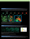
FLIP—Fluorescence Loss in Photobleaching
Fluorescence loss in photobleaching (FLIP) combines imaging with continuous bleaching of a specific region to observe the diffusion of a
target protein within a cell. The changes in the image over time make it possible to observe the location of structural bodies that inhibit
the diffusion of the molecule.
FRAP—Fluorescence Recovery after Photobleaching
Exposure of fluorescent-labeled target proteins to strong laser light causes their fluorescence to fade locally. Fluorescence recovery after
photobleaching (FRAP) is used to observe the gradual recovery of fluorescence intensity caused by protein diffusion from the area
surrounding the bleached region. By examining the resulting images, it is possible to characterize the diffusion speed of the molecule,
and the speed of binding and release between the molecule and cell structures.
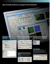
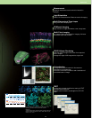
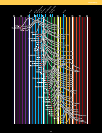
Example: Fluorescence recovery without interactions
If the protein can freely diffuse, the bleached region recovers
its fluorescence at a high speed due to Brownian motion.
Example: Fluorescence recovery with interactions
If the protein is strongly bound to a structure or forms part of a
large protein complex, the bleached region recovers its
fluorescence at a slower rate relative to the unbound state.
Time
Fluorescent intensity
Time
Fluorescent intensity
Specimen: HeLa cell, GFP (free), 488 nm excitation (multi-argon laser)
Image acquisition time: 100 ms/ bleach time: 100 s continuously, 405 nm bleaching
0
0
200
400
600
800
1,000
1,200
1,400
1,600
1,800
2,000
2,200
2,400
2,600
2,800
3,000
10,000 20,000 30,000 40,000 50,000
Time (ms)
60,000 70,000 80,000 90,000 100,000
Intensity
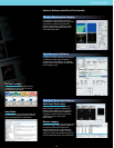
Specimen: Hippocampal neurons, Shank-GFP stain, 488 nm excitation (multi-argon laser)
Image acquisition time: 100 ms Bleach time: 80 ms, 488 nm excitation (Sapphire 488 laser)
Data courtesy of: Dr. Shigeo Okabe
Department of Anatomy and Cell Biology, Tokyo Medical and Dental University
0
250
300
350
400
450
500
550
600
650
750
700
10,000 20,000 30,000 40,000 50,000
Time (ms)
60,000 70,000 80,000
Intensity
17