


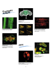


11
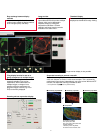
Lucifer Yellow: retina ganglion cell
TexasRed: dopamine-operated amacrine cell
Prof. Shigetada Nakanishi
Dept. of Biological Sciences,
Kyoto Univ. Faculty of Medicine
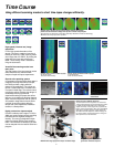
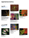
Applications Gallery
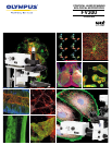
Morphology
Structure of PtK2 cell
Nucleus: DAPI (Blue)
Actin: FITC (Green)
Mitochondria: Mito Tracker (Red)
Microtubules: Cy5 (White)
Neuron
Rat tongue taste bud
DAPI: Nuclei
FITC: TrkB, high-affinity receptor for brain-derived
neurotrophic factor
Texas Red: Protein Gene Products
Pr. Shigeru Takami
Department of Anatomy,
School of Health Science,
Kyorin University
Lucifer yellow injected visual interneurons of
swallowtail butterfly
Extended focus is used for every 100µm on 383µm
Z-range image and displayed by overlapping pseudo colors
Mituyo Kinoshita, Pr. Kentaro Arikawa
Laboratory of Neuroethology,Graduate School of
Integrated Science, Yokohama City University
Purkinje cell in the rat cerebellum
FITC: vesicular GABA transporter VGAT
Cy3: vesicular glutamate transporter VGLUT1
Pr. Masahiko Watanabe
Department of Anatomy,
Hokkaido University School of Medicine
Mouse hippocampal neurons
GFP: postsynaptic density protein
Rhodamine-phalloidin: actin
Hippocampal neurons expressing a GFP-tagged
postsynaptic density protein were fixed and
stained with rhodamine-phalloidin to visualize the
localization of cytoplasmic actin filaments.
In dendrites, actin filaments are concentrated in
the postsynaptic sites.
Shigeo Okabe
Department of Anatomy and Cell Biology
Tokyo Medical and Dental University
Human Colon Crypt
Alexa 488 and To-Pro 3
Christine Anderson,Prof. Ray White's Laboratory,
Huntsman Cancer Institute, U. Utah