


The guidewire and injection lumens are separated to
ensure smoother passage of the guidewire. Additionally,
contrast media can be injected without removing the
guidewire. Two models are available — one with the
injection port located above the balloon and the other
with the injection port located below the balloon.
Triple-lumen design allows for
easy passage of the guidewire
The balloon catheter sheath is manufactured from a
special material that allows it for improved insertion
into the papilla, ensuring smoother guidewire passage,
and facilitating contrast injection. The tapered design
— 7 Fr. catheter at the proximal end tapers to 5 Fr. at
the distal end — enables an easy approach to the bile
duct and accommodates a 0.035" guidewire.
Easier cannulation
and tapered sheath design
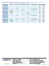
Multiple balloon sizing
Injection Lumen
0.035" Guidewire Lumen
Air Lumen
The balloon can be inflated to
one of three diameters— 8.5mm,
11.5mm, and 15mm. The balloon size can
be easily adjusted to suit the requirements
and conditions of each case so there is
no need to change catheters during
the procedure.
Reliable, Efficient Stone Extraction with the Multi-Size Balloon Design, Th
Manufactured from a Special Material That Allows for Easier Cannulation,
Tr iple Lumen, Multi Size Balloon with
Revolutionary V-System Exchange Capability for More
Precise Balloon Inflation and Efficient Stone Extraction
V-Marking
The exclusive V-Marking is located on the proximal side of the sheath. When
this marking reaches the channel port on the scope’s control section, it indicates
that the device tip has reached the distal end of the scope and the V-Groove
forceps elevator may be lowered. When withdrawing the device from the scope,
the same marking indicates when to raise the elevator to lock the guidewire.
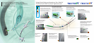
The V-System is a complete system that integrates Olympus endoscopes and EndoTherapy devices. The revolutionary
V-System design offers the option of guidewire manipulation by the physician or the assistant, allows easier exchange
of catheters, and enhances cannulation capability.
The V-Sheath allows the endoscopist complete device control or, if preferred,
device control may be given to the assistant. The unique device design allows
the guidewire sheath and injection sheath/handle to be separated. This forked
sheath design allows either the endoscopist or the assistant to control the device.
V- Sheath
The Innovative V-System Design Lets You Proceed
with Confidence and Efficiency
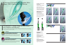
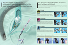
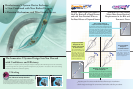
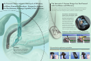
V-System device replacement procedure
C-Hook
The convenient C-Hook allows the device handle to be attached to the
endoscope’s control section, putting it within easy reach of the endoscopist.
With the device handle right at hand, the endoscopist can maneuver the
guidewire, inject contrast media, and manipulate the handle — all while keeping
a grip on the scope control section.
Indicates when to raise and lower
the V-Groove forceps elevator.
Device control by the endoscopist
or the assistant.
Confirm the position of the V-Marking
on the V-System EndoTherapy accessory.
Now endoscopists have the option to
manipulate guidewires and devices.
When the V-Marking is completely visible
above the instrument channel port, lift the
forceps elevator to lock the guidewire.
The guidewire is now locked
into the V-Groove.
Completely remove the device.