


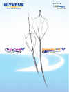

Exceptional Cutting Performance and Easy,
Fast Exchange Capability for Enhanced Efficiency
in ERCP Sphincterotomy
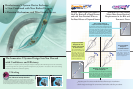
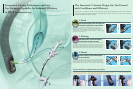
Distal marking on the sheath for
improved view field visibility
The distal marking on the sheath
clearly indicates both the center and
cutting position of the knife.
Injection lumen
Guidewire lumen
Cutting wire lumen
Sheath design for stable and
reliable cannulation
Designed to optimize insertion into the
scope, this sheath is narrower at the distal
end and thicker at the proximal end.
This improves handling and ensures smoother
insertion, while also providing excellent
cannulation capability into the papilla.
Easy identification
of ports
The guidewire port and the
injection port are easily
identified by symbols.
Unique Device Design and Attention to Every Detail of
The CleverCut2V and CleverCut3V Sphincterotomes
CleverCut coating enhances safety
Olympus’s signature CleverCut coating on the
proximal end of the cutting wire minimizes damage
to the surrounding tissue. In addition,
CleverCut Coating reduces the risk of electrical
contact between the wire and the endoscope.
The CleverCut3V wire, injection lumen and guidewire
lumen are arranged to allow easier orientation of the
cutting wire for effective sphincterotomy. Since the
injection lumen and the guidewire lumen are completely
separate, contrast media can be smoothly injected with
a guidewire in place.
The CleverCut3V offers excellent
orientation and smooth injection
Features that display
the icon on the top
are available with
CleverCut2V
double-lumen
models. Those
displaying the
bottom icon are
available with
CleverCut3V
triple-lumen models.
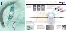
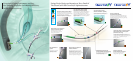
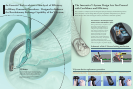
V-Marking
The exclusive V-Marking is located on the proximal side of the sheath. When
this marking reaches the channel port on the scope’s control section, it indicates
that the device tip has reached the distal end of the scope and the V-Groove
forceps elevator may be lowered. When withdrawing the device from the scope,
the same marking indicates when to raise the elevator to lock the guidewire.
The V-Sheath allows the endoscopist complete device control or, if preferred,
device control may be given to the assistant. The unique device design allows
the guidewire sheath and injection sheath/handle to be separated. This forked
sheath design allows either the endoscopist or the assistant to control the device.
V- Sheath
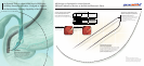
The Innovative V-System Design Lets You Proceed
with Confidence and Efficiency
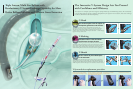
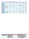
V-System device replacement procedure
C-Hook
The convenient C-Hook allows the device handle to be attached to the
endoscope’s control section, putting it within easy reach of the endoscopist.
With the device handle right at hand, the endoscopist can maneuver the
guidewire, inject contrast media, and manipulate the handle — all while keeping
a grip on the scope control section.
Indicates when to raise and lower
the V-Groove forceps elevator.
Device control by the endoscopist
or the assistant.
Confirm the position of the V-Marking
on the V-System EndoTherapy accessory.
Now endoscopists have the option to
manipulate guidewires and devices.
When the V-Marking is completely visible
above the instrument channel port, lift the
forceps elevator to lock the guidewire.
The guidewire is now locked
into the V-Groove.
Completely remove the device.
The V-System is a complete system that integrates Olympus endoscopes and EndoTherapy devices. The revolutionary
V-System design offers the option of guidewire manipulation by the physician or the assistant, allows easier exchange of
catheters, and enhances cannulation capability.