


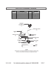

SKU 54949 For technical questions, please call 1-800-444-3353 PAGE 5
8. Diaphragm: The Diaphragm (9) is placed in the light path to alter the amount of
light that reaches the condenser within the Microscope (for enhancing contrast in
the image). (See Assy. Diagram.)
9. Base: The Base (12) supports the weight of all of the Microscope parts.
(See Assy. Diagram.)
Specimen Preparation:
1. When observing a specimen by transmitted light, light must pass through the
specimen in order to form an image. The thicker the specimen, the less light
passes through. The less light that passes through, the darker the image.
2. The specimens must be thin (0.1 to 0.5mm).
3. Many specimens must be cut into thin sections before observation. Specimens
such as rock or semiconductors are too thick to be sectioned and observed by
transmitted light, so they are observed by the light reflected from their surfaces.
Microscopic Terms:
1. Depth of Field: The vertical distance, from above to below the focal plane, that
yields an acceptable image.
2. Field of View: The area of the specimen that can be seen through the Micro-
scope with a given Objective Lens (5).
3. Focal Length: The distance required for an Objective Lens (5) to bring the light
to a focus (usually measured in microns).
4. Focal Point/Focus: The point at which the light from an Objective Lens (5)
comes together.
5. Magnification: The product of the magnifying powers of the Objective Lenses
(5) and Zoom Eyepiece (1). The numbers marked on the Objective Lenses
indicates how many times the specimen on the Slide (7) is being magnified. The
Objective Lenses on this Microscope allow a specimen to be magnified from
eight (8X) to twenty (20X) times its actual size.