


FF
C-mount Camera
H
F
F
A
B
C
D
A
B
C
D
Y-TV
TV Tube
Y-TB
Binocular
Tube B
Y-TF
Trinocular
Tube FUW
Y-TT
Trinocular
Tube TUW
C-TE
Ergonomic
Binocular Tube
C-TEP
DSC Port
DS AC Adapter
Li-ion
Battery
EN-EL1
Darkfield
* Darkfield*
Condenser
(oil)
Condenser
(dry)
C-C
/Aplanat
Condenser
C-C
AchromatAchromat
Condenser
Swing-out
Condenser
C-C
Abbe
Condenser2-100x
LWD
Achromat
Condenser
C-C
Achromat
Swing-out
Condenser
1-100x
C-C
*
Phase
Condenser
C-C
Low Power
CondenserContrast
Attachment
Epi-fl
Filter Cubes
D
C-SP*
Simple
Polarizer
C-TP
*
Polarizer with
First-order Red
Tint Plate
C-ER Eye-level Riser
C-ISA Intermediate Tube
*
w/Simple Analyzer
C-IA Intermediate Tube*
w/Analyzer
Y-IDP Double Port (45-55 / 0-100, 0-100)
Y-IM Magnification Module
Y-IDT Drawing Tube
Y-THF Teaching Unit Face to Face
*
Y-THS Teaching Unit Side by Side B*
Y-THPS Support for Side by Side
Y-THM Main Teaching Unit
*
Y-THR Teaching Unit Side by Side A
Y-THP Pointer Unit
Y-THA AC Adapter
B
A
CFI60
Objective Lens
J-CY
Cytodiagnostic
Unit
C-HS
Hand Switch
Cable Set
for 55i &
Cytodiagnostic
Unit
C-SR
Mechanical
Stage
C-N
Sextuple
Nosepiece
H H
F
E
CFI
12.5x
CFI
10x
CFI
10x M
CFI
15x
CFI UW
10x
CFI UW
10x M
C-CT
Centering
Telescope
C-FC
Epi-Fl
Collector Lens
Quartz Epi-FI
Collector Lens
C-FC
Epi-Fl
Collector Lens
C-FC
Epi-Fl
Collector Lens
Mercury Lamp
Socket S 100W
Xe Lamp
Socket 75W
Halogen Lamp
Socket 100W/HMX
Hg Lamphouse
HMX-3B
Hg Lamphouse
HMX-4B
Xe Lamphouse
HMX-4
HMX Lamphouse
N
C-SHG1
Power Supply for HG100W
Xenon Power Supply 75W
UN2 Transformer 100W
Camera Mounts
Eyepiece Tubes
Nosepieces
Polarizers
Condensers
Intermediate Modules
Stages / Specimen Holders
Lamphouses
Eyepieces
N
TE-AT Double Lanphouse
Adapter
N
N
N
C-HL2
Specimen Holder Specimen Holder Specimen Holder
(2 slides: Left)
C-HR2
(2 slides: Right)
C-HC1
(1 slide)
GG
G
Y-QT
Quadrocular
Adapter
C
C-CEL Expander Lens
*1: Use the dedicated 0.45X adapter for the double port sub-port.
F
E
F
E
ENG-mount Camera C-mount Camera
ENG-mount
TV Adapter*
1
0.6x
ENG-mount
TV Adapter
Adapter
Relay
Lens
1x
ENG-mount
Zooming
Adapter
Zooming
C-mount
TV Zoom
Lens
Relay
Lens
1x
C-mount
TV Adapter
C-mount TV
Adapter
0.35x
0.38x
0.45x
0.6x
C-mount
TV Adapter A
C-mount
TV Adapter
VM2.5x
C-mount
TV Adapter
VM4x
V-T
Photo
Adapter
FX-III Series
Photomicrographic System
Projection Lens
PLI 2x
PLI 2.5x
PLI 4x
PLI 5x
C-0.7x
DXM Relay Lens
G
1110
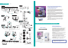
System Diagram
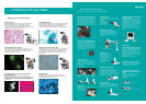
Nikon offers various options to best suit your digital-imaging needs
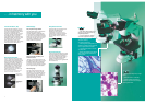
Digital microscope
The new Plan Apo VC objectives and unique “fly-eye” optics guarantee uniform
brightness over the whole view field and unparalleled resolution to the
peripheries of the image. These remarkable achievements, which take digital
imaging to new heights, are the result of Nikon’s breakthroughs in optical
technologies and precision engineering.
—Hi S/N Fluorescence System. The universal epi-fluorescence illuminator and
digital-imaging head incorporate Nikon’s unique Hi S/N Fluorescence System,
which employs a “Noise Terminator” to achieve unparalleled contrast with
excellent S/N ratios. An optional “Excitation Balancer” allows specific wavelengths
to be emphasized in multi-stained specimens.
— Digital-Imaging Head. This creates an optimum digital-imaging system that
enables fluorescence imaging with outstanding results. When the DS-5M-L1 digital
camera is mounted to this head, observation data such as magnification and
fluorescence filters in use is automatically detected and can be saved together
with the image file.
— New DIC System. A perfect balance of high resolution and high contrast is
possible, even at low magnifications. Three types of DIC prisms are available:
standard, high-contrast, and high-resolution.
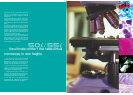
Built around an optimum digital-imaging platform,
the 80i can visualize weakly fluorescing molecules with
much higher brightness and contrast,
while reducing background noise.
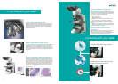
An all-in-one microscope
that can be operated by mouse
The COOLSCOPE is revolutionary in every respect, from its looks
to its functionality and ease of use. After loading the slide glass
preparation onto the tray, users need only click the mouse to
operate the microscope. Observations, image recording and
networking can all be done with a single unit of the COOLSCOPE—
no need for a PC. The popular image of a microscope has
undergone a major metamorphosis.
— Motorized operation enables brightfield observations through the
monitor’s GUI.
— Simultaneous display of both enlargements (micro) of specimen
portions and whole image (macro) on a single screen.
— The point of interest can be easily moved to the center by simply
clicking the point in the micro image. In the macro image, the
selected point is enlarged and displayed in the micro image.
— Instant recall of previous observations can be achieved by clicking
the numbered button.
— Remote operation* and viewing of images is possible through
networked computers anywhere.
* Some operations are limited
Please visit our dedicated website, where you will find
extensive information on the uses and operation of the
COOLSCOPE.
www.coolscope.com
Advanced research microscope
*: 50i model.